1. Introduction to Cellular Optical Barcoding |
1.1 Cellular ¡°optical barcoding¡± is a revolutionary technology that allows researchers to assign unique, optically readable identifiers to individual cells through the use of microscopic light-emitting particles known as laser particles or micro-lasers. These particles, once introduced into cells, emit distinct optical signatures¡ªsuch as specific laser wavelengths, emission spectra, or resonance frequencies¡ªthat can be detected and decoded using optical microscopes or spectroscopic instruments. |
1.2 This system effectively functions as a barcode system at the cellular level: instead of using printed black and white stripes or 2D patterns, optical barcoding encodes information through light characteristics such as color, wavelength spacing, intensity, and polarization. Each cell receives a unique optical ¡°fingerprint,¡± which can be continuously monitored over long time periods without interfering with cell viability or function. |
1.3 The significance of this technology lies in its ability to address a fundamental challenge in cell biology and biomedicine: the ability to track and identify millions of individual living cells over time, in complex biological systems, with high resolution and minimal disturbance. Previous labeling methods¡ªsuch as fluorescent dyes, genetic reporters, or nanoparticle tags¡ªeither suffer from photobleaching, instability, or limited distinguishable combinations. Optical barcoding using laser particles overcomes these barriers, allowing long-term, non-destructive, and scalable cell tracking. |
1.4 The development of cellular optical barcoding represents a convergence of several advanced scientific domains: nanophotonics, materials science, bioengineering, and single-cell biology. It embodies the idea of merging photonics with life sciences¡ªusing light not merely as a visualization tool, but as a carrier of encoded information within living systems. |
|
2. The Concept and Principle of Optical Barcoding |
2.1 In its most fundamental form, optical barcoding refers to the generation of a unique light-emission signature that can serve as an identifying code. In biological systems, this signature must satisfy several conditions: it should be highly stable over time, uniquely identifiable among many cells, non-toxic, and detectable through standard optical or spectroscopic instruments. |
2.2 The term ¡°barcode¡± here is metaphorical. Instead of spatial patterns, optical barcodes use spectral patterns. Each barcode corresponds to a specific combination of emission wavelengths or spectral lines. The more finely these spectral characteristics can be controlled, the greater the number of unique barcodes that can be generated. |
2.3 Laser particles serve as the physical embodiment of these optical barcodes. Each laser particle functions as a miniature resonant cavity capable of generating coherent light when excited. The resonant wavelength depends on the cavity size, refractive index, and shape. Therefore, by fabricating laser particles of slightly varying diameters or material compositions, researchers can create a virtually limitless array of emission spectra. |
2.4 When such particles are internalized by cells¡ªtypically via endocytosis¡ªthey act as optical ¡°ID tags.¡± Upon illumination with an external light source, each tag emits its unique spectral fingerprint, allowing identification of the cell in which it resides. This enables tracking of individual cells through multiple generations, within tissues, or under dynamic biological conditions. |
2.5 The decoding process resembles spectral analysis. Using spectrometers or hyperspectral imaging systems, researchers can read the emitted wavelengths and assign each to a known code, thereby determining the identity of each cell. This process is analogous to scanning a barcode in retail logistics, except here the ¡°scanner¡± is an optical microscope and the ¡°product¡± is a living cell. |
|
3. Historical Background and Scientific Motivation |
3.1 The motivation for developing cellular optical barcoding originates from limitations in existing cell labeling and tracking methods. Traditional approaches include: |
(1) Fluorescent dyes and proteins, which offer multiple colors but are limited by overlapping spectra and photobleaching. |
(2) DNA barcoding, which uses genetic sequences as identifiers but requires destructive sequencing to read. |
(3) Magnetic or metallic nanoparticles, which are durable but provide limited multiplexing capacity and may interfere with cellular functions. |
3.2 As single-cell biology advanced¡ªespecially with the rise of single-cell RNA sequencing and live-cell imaging¡ªscientists realized that to fully understand complex systems such as tumor evolution, stem cell differentiation, or immune response, they needed a way to track thousands or even millions of cells individually over long timescales. |
3.3 Around the 2010s, researchers in photonics and nanotechnology began exploring micro- and nano-lasers as optical probes. Microdisk and microsphere lasers, originally developed for optical communication and sensing, demonstrated highly stable and tunable emission wavelengths. This inspired the concept that if such micro-lasers could be miniaturized, made biocompatible, and introduced into living cells, they could act as permanent optical identifiers. |
3.4 Early proof-of-concept studies demonstrated that whispering-gallery-mode (WGM) resonators¡ªtiny circular structures that trap light through total internal reflection¡ªcould generate stable, narrowband laser emission. Each resonator¡¯s emission wavelength was directly related to its size. By controlling the diameter during fabrication, unique optical codes could be defined. |
3.5 This realization laid the foundation for what is now known as cellular optical barcoding: an approach that uses photonic microstructures as encoded identifiers inside biological systems. |
|
4. Fundamental Physics of Laser Particles |
4.1 Laser particles used in cellular optical barcoding are typically micrometer-scale spherical or disk-shaped cavities that confine light through internal reflection, forming standing optical waves known as whispering-gallery modes (WGMs). These modes occur when light circulates around the cavity perimeter, reinforcing itself through constructive interference. |
4.2 The resonance condition for a WGM cavity is given by the relationship between cavity circumference and wavelength: |
m¦Ë = 2¦Ðr n_eff, |
where m is the mode number, ¦Ë the resonant wavelength, r the radius, and n_eff the effective refractive index. Slight variations in r lead to precise shifts in ¦Ë, which can be measured with high accuracy. |
4.3 When optically pumped¡ªtypically by an external pulsed or continuous-wave laser¡ªthe embedded dye or semiconductor gain medium within the particle amplifies light at the resonant wavelength, resulting in coherent laser emission. This emission can be extremely narrow (on the order of 0.1 nm linewidth) and stable against photobleaching, in stark contrast to traditional fluorescence signals. |
4.4 Because the resonance wavelength is determined geometrically, not chemically, the barcode is intrinsic and does not degrade with time. This geometric encoding makes it ideal for long-term tracking. |
4.5 The output signal can be detected by spectrometers or hyperspectral cameras, which record the emission wavelength distribution. Each unique combination of resonant peaks forms a distinct optical barcode that can be decoded computationally. |
4.6 Additionally, multimode emission or combinations of multiple particles per cell can exponentially increase the total number of available unique codes¡ªreaching millions or more theoretically. |
5. Fabrication of Laser Particles |
5.1 The fabrication of laser particles suitable for biological use requires precise control of dimensions, optical properties, and biocompatibility. Several microfabrication methods are used: |
5.2 Semiconductor microdisks: Fabricated using lithography and etching from materials like InGaAsP or GaAs. These produce well-defined resonant modes but require passivation layers for biological use. |
5.3 Polymer microspheres: Created by emulsion or microfluidic droplet formation. Polymers such as polystyrene or polymethyl methacrylate (PMMA) can be doped with laser dyes (e.g., Rhodamine, Coumarin) to form biocompatible gain media. |
5.4 Silica microbeads: Formed via sol-gel or flame hydrolysis methods. They are optically stable and can be coated with thin dye-doped polymer shells for gain. |
5.5 Dielectric microshell lasers: Consist of a core-shell structure with different refractive indices to enhance confinement and reduce threshold. |
5.6 The fabrication process involves controlling particle diameter at nanometer precision. For example, a 1 nm change in radius can shift the resonant wavelength by approximately 0.6 nm, providing fine spectral tuning. |
5.7 To achieve biocompatibility, particles are typically coated with silica, PEG (polyethylene glycol), or lipid bilayers to prevent cytotoxicity and aggregation. Surface functionalization also enables targeted uptake by specific cell types. |
5.8 Quality control involves verifying spectral output uniformity, emission stability, and particle integrity under biological conditions such as in saline or cell culture media. |
6. Introduction into Cells |
6.1 The introduction of laser particles into living cells is a critical step in creating optical barcodes. Several methods are employed depending on cell type and desired localization: |
6.2 Endocytosis: Cells naturally internalize nanoparticles via endocytosis. By adjusting particle surface charge and size (typically 2¨C10 ?m), uptake can occur spontaneously. |
6.3 Microinjection: For precise placement or larger particles, microinjection directly inserts a laser particle into the cytoplasm using a microneedle under a microscope. |
6.4 Electroporation: Brief electric pulses temporarily permeabilize cell membranes, allowing particles to enter without significant damage. |
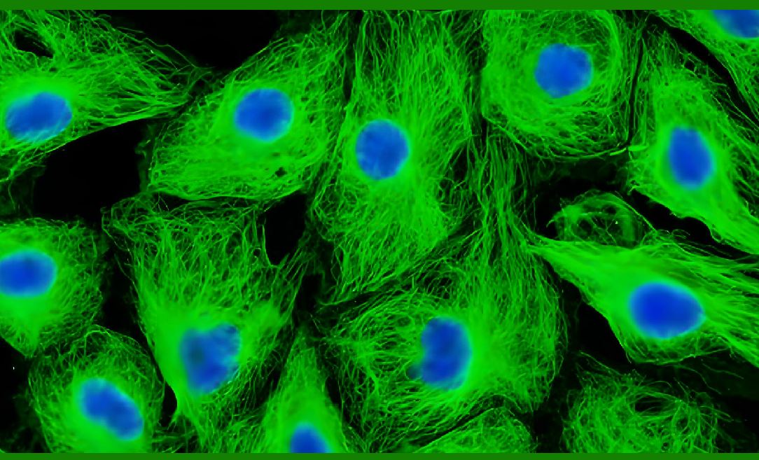
6.5 Once inside, particles typically localize in the cytoplasm and remain there without entering the nucleus. Studies have shown that cells can divide normally, passing the particles to daughter cells, which allows lineage tracing over many generations. |
6.6 The non-toxic coatings and low excitation intensities used ensure minimal photothermal or mechanical disruption to cellular physiology. |
6.7 Stability studies show that internalized laser particles remain optically stable for weeks to months, even under continuous cell culture conditions, enabling longitudinal studies of dynamic biological processes. |
|
7. Reading and Decoding of Optical Barcodes |
7.1 To read optical barcodes, the cells are illuminated with a pump laser tuned to excite the particle¡¯s gain medium (often in the visible or near-infrared region). |
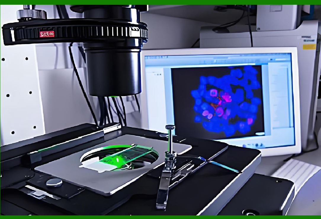
7.2 The emitted light is collected through a microscope objective and analyzed using a spectrometer. The emission spectrum shows sharp peaks corresponding to the resonant wavelengths. |
7.3 Each unique emission spectrum acts as the cell¡¯s identifier. By mapping these spectra to a database, one can track the position, behavior, or state of specific cells over time. |
7.4 For high-throughput operation, hyperspectral imaging systems or multiplexed microfluidic setups can simultaneously record the spectra of thousands of cells. |
7.5 Computational decoding algorithms perform spectral clustering, peak fitting, and wavelength mapping to assign each emission signature to a barcode ID. |
7.6 The precision of wavelength identification allows multiplexing of millions of cells, far exceeding the capabilities of traditional fluorescent labeling, which is typically limited to fewer than ten distinct colors. |
7.7 In advanced implementations, multiple laser particles with different wavelengths can be inserted into one cell, producing combinatorial codes. Each cell¡¯s barcode becomes a unique vector of emission wavelengths. |
7.8 These optical codes can then be correlated with cellular parameters such as migration trajectories, division rates, or gene expression profiles, allowing dynamic, high-dimensional analysis. |
8. Biological Compatibility and Safety |
8.1 One of the most important aspects of cellular optical barcoding is ensuring that laser particles do not alter cellular physiology. |
8.2 Numerous biocompatibility studies have demonstrated that properly coated micro-lasers do not affect cell viability, proliferation, or gene expression. Coatings like silica and PEG reduce immune responses and prevent protein adsorption. |
8.3 The excitation power used to induce lasing is low enough to avoid heating or photodamage. Typically, a few microjoules per pulse suffice to trigger emission, far below cytotoxic levels. |
8.4 Cytoplasmic localization ensures that the nucleus remains unaffected, minimizing interference with genetic functions. |
8.5 Long-term imaging experiments have shown that cells containing laser particles continue normal activities such as migration, differentiation, and cytokinesis for weeks. |
8.6 In vivo experiments¡ªe.g., laser-particle-labeled cells implanted in animal models¡ªshow successful tracking of cellular migration and tissue integration without adverse immune reactions. |
|
9. Scalability and Multiplexing Capacity |
9.1 The number of distinct optical barcodes achievable depends primarily on how finely laser wavelengths can be distinguished. |
9.2 Typical WGM lasers have linewidths of 0.1 nm and can be tuned over a range of hundreds of nanometers. Thus, even a modest 200 nm spectral range theoretically allows over 2,000 distinct single-particle codes. |
9.3 Combinatorial encoding using multiple particles per cell exponentially expands the code space. For example, two particles yield millions of combinations. |
9.4 Additionally, multimode lasers or hybrid materials can produce multiple peaks per particle, further increasing diversity. |
9.5 This scalability allows labeling of enormous cell populations¡ªon the order of millions¡ªeach with a distinct optical signature. |
9.6 The capability surpasses any previous optical labeling method, providing the foundation for large-scale cellular lineage and interaction studies. |
10. Applications in Biomedical Research |
10.1 The core purpose of cellular optical barcoding is to enable detailed tracking and analysis of individual cells within complex biological systems. The following are major application areas: |
10.2 Tumor Cell Tracking: |
Cancer progression involves heterogeneous populations of cells with varying proliferation and metastasis abilities. Optical barcoding enables tracking of each tumor cell¡¯s behavior within living tissue over time. Researchers can observe how specific clones dominate or regress, identify metastatic precursors, and evaluate drug responses at single-cell resolution. |
10.3 Stem Cell Lineage Analysis: |
Stem cells divide asymmetrically and differentiate into multiple lineages. By assigning each stem cell a unique optical code, researchers can reconstruct lineage trees over long periods, revealing developmental hierarchies and fate decisions. |
10.4 Immunology and Cell Migration Studies: |
Tracking immune cells as they patrol tissues or infiltrate tumors is critical for understanding immune responses. Optical barcoding allows simultaneous observation of thousands of immune cells, distinguishing between different activation states or migration patterns. |
10.5 Drug Screening: |
High-throughput pharmacological assays can use optical barcoded cells to study how individual cells respond differently to the same drug, identifying resistant subpopulations in heterogeneous samples. |
10.6 Neuroscience: |
In neural cultures, optical barcodes can label individual neurons, enabling long-term monitoring of network formation, synaptic connections, and activity dynamics. |
10.7 Regenerative Medicine: |
During tissue regeneration, optical barcoded cells can reveal which cells contribute most effectively to repair, helping optimize therapeutic strategies. |
10.8 Single-cell Omics Integration: |
Optical barcodes can be combined with single-cell RNA sequencing or proteomics. Cells can first be tracked optically, then isolated and sequenced. The optical code serves as a persistent link between live behavior and molecular profile. |
|
11. Clinical Diagnostic and Therapeutic Potentials |
11.1 Beyond laboratory research, optical barcoding has significant potential in clinical diagnostics and therapeutic monitoring. |
11.2 Personalized Oncology: |
By labeling patient-derived tumor cells, clinicians can track their proliferation and migration in vitro or in vivo, testing personalized therapies on uniquely identified subclones. |
11.3 Cell-based Therapies: |
Stem cells or engineered immune cells (e.g., CAR-T cells) used for therapy can be optical-barcoded before administration, allowing clinicians to monitor their distribution, survival, and activity in patients. |
11.4 Circulating Tumor Cell Detection: |
Optical barcodes can identify specific circulating tumor cells in blood samples, enabling early cancer detection and prognosis monitoring. |
11.5 Intraoperative Tracking: |
Surgeons could potentially visualize optical barcoded cells in real time using spectroscopic endoscopes to ensure precise tumor removal or confirm engraftment of therapeutic cells. |
12. Advantages over Traditional Labeling Methods |
12.1 High Multiplexing Capacity ¨C Millions of unique codes possible versus fewer than ten with fluorescent dyes. |
12.2 Non-destructive Readout ¨C Optical decoding does not require cell lysis or genetic sequencing. |
12.3 Long-term Stability ¨C Laser emission remains stable for months; dyes photobleach within hours. |
12.4 High Signal-to-noise Ratio ¨C Coherent laser light offers stronger contrast than fluorescence. |
12.5 Compatibility with Live Imaging ¨C Enables real-time observation without altering cell behavior. |
12.6 Scalable Fabrication ¨C Microfluidic synthesis can mass-produce uniform particles. |
12.7 Biocompatibility ¨C Coatings prevent toxicity and immune responses. |
|
13. Technical Challenges and Limitations |
13.1 Despite its potential, cellular optical barcoding faces several technical and practical challenges. |
13.2 Fabrication Uniformity ¨C Producing millions of laser particles with precisely controlled sizes remains difficult at industrial scales. Small size deviations can lead to wavelength overlaps. |
13.3 Detection Throughput ¨C Current spectral imaging systems are limited in speed; reading millions of barcodes simultaneously remains a bottleneck. |
13.4 Intracellular Position Effects ¨C Particle position within the cell can slightly affect emission spectra due to local refractive index changes. |
13.5 Cell Division Dilution ¨C Over many generations, daughter cells may not inherit equal numbers of particles, leading to barcode loss in some lineages. |
13.6 In Vivo Optical Access ¨C Deep-tissue detection is limited by light scattering; near-infrared lasers may alleviate this but require further optimization. |
13.7 Regulatory Approval ¨C Clinical translation demands extensive safety testing and biocompatibility validation. |
14. Advances in Decoding and Data Analysis |
14.1 As optical barcoding produces large spectral datasets, computational methods play a vital role in decoding and analysis. |
14.2 Machine Learning Algorithms can classify spectral fingerprints with high accuracy, distinguishing subtle wavelength differences even under noise. |
14.3 Spectral Unmixing Techniques separate overlapping peaks in dense datasets, ensuring reliable identification. |
14.4 Automated Cell Tracking Software integrates optical barcode data with spatial coordinates from live imaging, reconstructing dynamic cell trajectories. |
14.5 Big Data Integration links optical identifiers with omics datasets, enabling multimodal cellular analysis across time, space, and molecular dimensions. |
14.6 Cloud-based frameworks now allow collaborative barcode tracking experiments across research centers. |

|
15. Future Development Trends |
15.1 Miniaturization: Advances in nanofabrication will yield smaller laser particles (<1 ?m), allowing insertion even into small cells such as lymphocytes. |
15.2 Tunable Emission Materials: New gain media such as perovskites or quantum dots will expand spectral tunability and improve efficiency. |
15.3 Near-infrared Lasers for Deep Imaging: Shifting to NIR wavelengths will permit in vivo imaging through tissue layers. |
15.4 Hybrid Barcoding Systems: Combining optical barcodes with genetic or chemical labels will provide multidimensional identifiers. |
15.5 Real-time In Vivo Tracking Devices: Portable spectroscopic microscopes may enable live monitoring of barcoded cells in animal models or patients. |
15.6 Standardization of Code Libraries: Future research may define standardized optical barcode ¡°libraries¡± analogous to genetic sequence databases, ensuring reproducibility across laboratories. |
15.7 Integration with AI: Artificial intelligence will enhance spectral decoding, anomaly detection, and cell behavior prediction from large-scale barcode datasets. |
16. Ethical and Regulatory Considerations |
16.1 As with any technology involving modification of living cells, ethical guidelines must be considered. |
16.2 Data Privacy: When used for patient-derived cells, barcode data should be anonymized and secured. |
16.3 In Vivo Applications: Introducing foreign materials into patients requires safety evaluation, particularly regarding long-term biostability and clearance. |
16.4 Environmental Impact: Disposal of nanoparticle-containing waste must comply with biosafety regulations. |
16.5 Equitable Access: Advanced tracking technologies should be accessible to research communities globally, avoiding inequality in biomedical progress. |

|
17. Potential Industrial and Commercial Uses |
17.1 Although primarily a research tool today, cellular optical barcoding could evolve into a commercial platform. |
17.2 Pharmaceutical Screening Services: Companies may use barcoded cells for drug efficacy and toxicity screening with unprecedented precision. |
17.3 Clinical Diagnostic Kits: Optical barcode-based cytology assays could provide unique identifiers for rare cells in samples such as blood or tissue biopsies. |
17.4 Biosensor Development: Laser particles could serve as high-sensitivity biosensors for detecting molecular changes inside living cells. |
17.5 Educational and Analytical Tools: Academic institutions might adopt optical barcoding to teach photonics¨Cbiology integration. |
18. Comparative Perspective with DNA Barcoding |
18.1 DNA barcoding uses genetic sequences as identifiers, while optical barcoding uses light. Both aim at tracking individual cells but differ fundamentally. |
18.2 DNA barcoding offers enormous encoding capacity but requires destructive sequencing. Optical barcoding offers real-time, non-destructive readout. |
18.3 Optical barcodes are re-readable continuously, whereas DNA barcodes are readable only once after cell lysis. |
18.4 However, DNA barcoding integrates easily with molecular biology workflows, while optical barcoding requires specialized optical systems. |
18.5 The two can complement each other: optical barcodes track live dynamics, and DNA barcodes record lineage history retrospectively. |
19. Example Experimental Workflows |
19.1 A typical optical barcoding experiment proceeds as follows: |
(1) Fabricate or purchase laser particles with defined diameter distributions. |
(2) Coat particles with biocompatible layers and functionalize surfaces if needed. |
(3) Introduce particles into cultured cells via endocytosis or microinjection. |
(4) Verify intracellular localization via confocal microscopy. |
(5) Excite the particles and record emission spectra. |
(6) Assign spectral peaks to barcode IDs. |
(7) Monitor cell behavior over time using live-cell imaging. |
(8) Decode spectral data periodically to track each cell¡¯s identity and position. |
(9) Analyze correlations between optical codes and biological parameters. |
19.2 This workflow can be automated for high-throughput cell-tracking platforms. |
|
20. Long-term Vision: Photonic Identity for Cells |
20.1 The ultimate vision of cellular optical barcoding is to provide each living cell with a unique, persistent photonic identity¡ªanalogous to a digital fingerprint for biological systems. |
20.2 In future biomedical ecosystems, cells could be optically tagged before therapeutic use, and their behavior tracked throughout treatment, ensuring efficacy and safety. |
20.3 In fundamental research, scientists could construct complete dynamic maps of cellular interactions in tissues, tracing every cell¡¯s lineage, migration, and communication pathways. |
20.4 Combined with machine learning, these datasets could enable predictive models of disease progression and tissue development. |
20.5 Thus, cellular optical barcoding is not merely a labeling technology¡ªit represents a paradigm shift in how we observe and understand life at the single-cell level through the language of light. |
21. Photonic Design Optimization of Laser Particles |
21.1 The effectiveness of cellular optical barcoding fundamentally depends on the photonic design of the micro-lasers themselves. Each particle¡¯s emission characteristics¡ªwavelength, linewidth, threshold, and stability¡ªare determined by its geometry, material composition, and refractive environment. |
21.2 Whispering-Gallery-Mode (WGM) Resonators are the most common structure used for these optical barcodes. Their circular geometry allows photons to circulate multiple times around the circumference via total internal reflection, forming resonant modes that amplify specific wavelengths. |
21.3 Optimization involves balancing several parameters: |
(1) High Q-factor (quality factor) to achieve narrow spectral peaks. |
(2) Low lasing threshold to reduce required pump power. |
(3) Mechanical and chemical stability for biological conditions. |
21.4 A high Q-factor ensures that the resonator stores light efficiently, producing sharper and more distinguishable emission peaks. Typical Q-factors for polymer microsphere lasers reach 10?¨C10?, resulting in linewidths of less than 0.1 nm. |
21.5 Reducing the lasing threshold minimizes the energy needed to induce emission. This is crucial inside living cells where excessive optical power could cause heating or phototoxic effects. Thresholds below 1 ¦ÌJ per pulse are generally acceptable for biological imaging. |
21.6 Refractive Index Contrast between the resonator and surrounding medium (cytoplasm, typically n¡Ö1.36¨C1.38) determines mode confinement. Materials such as polystyrene (n¡Ö1.59) or semiconductor materials (n¡Ö3.4 for GaAs) provide strong confinement, enabling stable operation. |
21.7 Size Control is critical for encoding uniqueness. Each resonator¡¯s radius corresponds directly to a specific emission wavelength. Advanced lithographic or droplet-based methods can control diameters with sub-nanometer precision, ensuring reproducible spectral spacing. |
21.8 Thermal and Mechanical Stability are achieved by selecting materials resistant to swelling or degradation in aqueous media. Silica coatings prevent dye leaching and minimize refractive index fluctuations. |
21.9 Gain Medium Selection determines emission color range. Organic laser dyes offer tunability but may bleach; quantum dots or semiconductor materials provide robust, stable emission. Hybrid systems¡ªsuch as dye-doped polymer cores with quantum-dot shells¡ªoffer enhanced durability. |

|
22. Microfabrication Techniques and Engineering Control |
22.1 Several engineering methods exist to fabricate laser particles at high precision and throughput. |
22.2 Lithography-Based Fabrication uses semiconductor processes. Thin films of GaAs or InGaAsP are patterned into microdisks, then released into suspension. These provide precise control over size but are expensive and yield smaller quantities. |
22.3 Emulsion Droplet Synthesis creates polymer or dye-doped microspheres through fluid mixing in microfluidic channels. By adjusting flow rates and surfactant concentrations, particle diameters can be tuned continuously from hundreds of nanometers to tens of micrometers. |
22.4 Microfluidic Flow-Focusing Devices allow continuous production of uniform droplets at kilohertz frequencies. Real-time optical feedback systems can monitor emission spectra during production, ensuring spectral uniformity. |
22.5 Layer-by-Layer Coating provides surface functionalization. Alternating deposition of silica and PEG layers can tune biocompatibility, hydrophilicity, and optical boundary properties. |
22.6 Self-Assembly Approaches employ colloidal chemistry to grow uniform particles from solution without complex instrumentation. |
22.7 Dielectric Multilayer Cavities¡ªsuch as distributed Bragg reflector (DBR) microspheres¡ªuse alternating high- and low-index layers to confine light vertically, expanding wavelength control. |
22.8 Post-Fabrication Sorting employs optical tweezers or flow cytometry to separate particles by emission wavelength, creating well-defined spectral bins. |
22.9 Advances in additive manufacturing and nanoscale 3D printing now enable custom particle geometries¡ªellipsoidal, toroidal, or photonic-crystal-based¡ªto engineer emission polarization or angular dependence for additional barcode dimensions. |
23. Integration with Cellular and Molecular Biology |
23.1 Successful implementation of optical barcoding requires seamless integration with cellular environments. |
23.2 Surface Chemistry plays a decisive role. Particle surfaces can be functionalized with peptides, antibodies, or aptamers to target specific cell types. |
23.3 Controlled Uptake Kinetics can be tuned by manipulating particle charge (e.g., cationic coatings promote endocytosis) and by using specific cell-penetrating peptides. |
23.4 Intracellular Stability is maintained by avoiding lysosomal degradation. PEG coatings and neutral surface charges reduce endosomal trapping, allowing free cytoplasmic residence. |
23.5 Intracellular Localization Control is sometimes desirable. For example, attaching nuclear localization sequences can guide particles near the nucleus to monitor nuclear events, while mitochondrial-targeting peptides direct them to energy centers for metabolic tracking. |
23.6 Functional Compatibility ensures that laser emission remains unaffected by biochemical changes such as pH shifts or ionic strength variations. Silica shells act as protective barriers. |
23.7 Coexistence with Fluorescent Reporters: Optical barcodes emit narrow spectral lines that minimally overlap with conventional fluorophores, allowing simultaneous fluorescence imaging of cellular activity and barcode tracking. |
23.8 Cell Division Tracking: During mitosis, laser particles partition between daughter cells. Monitoring barcode inheritance allows construction of lineage trees, revealing asymmetric divisions and differentiation pathways. |

|
24. Large-Scale Data Collection and Automation |
24.1 As experiments expand to thousands or millions of cells, automation becomes essential. |
24.2 High-Content Microscopy Systems integrate automated stage movement, laser excitation, and spectral detection to scan large populations rapidly. |
24.3 Hyperspectral Imaging Cameras capture full spectral information for every pixel, enabling simultaneous decoding of thousands of barcodes per frame. |
24.4 Microfluidic Flow Systems can pass cells through laser interrogation zones at hundreds per second. Each cell¡¯s emission spectrum is recorded and tagged with spatial or temporal coordinates. |
24.5 Spectral Database Construction is necessary to map each emission pattern to a unique cell ID. Databases may contain millions of entries, requiring efficient indexing algorithms. |
24.6 Automated Spectral Matching Algorithms compare recorded spectra to known references using peak matching, cross-correlation, or machine-learning classifiers. |
24.7 Cloud-Based Processing Pipelines distribute decoding across multiple servers, enabling real-time tracking of large datasets. |
24.8 Visualization Interfaces render cell trajectories in 3D over time, integrating optical barcode identity with cell morphology and activity data. |
25. Case Study 1: Tumor Clonal Dynamics |
25.1 Cancer tumors are composed of heterogeneous subpopulations of cells, each with distinct genetic and phenotypic characteristics. Understanding how these subpopulations evolve under treatment or within microenvironments is a central challenge in oncology. |
25.2 In a landmark study, researchers labeled individual cancer cells with unique optical barcodes using dye-doped micro-lasers. |
25.3 The labeled cells were implanted into animal models. Over time, spectral imaging tracked the movement and proliferation of each subclone. |
25.4 By correlating emission spectra with tumor location and time, scientists reconstructed clonal expansion maps, identifying aggressive clones that dominated metastasis. |
25.5 The ability to trace the lineage and migration of thousands of cells simultaneously provided insights that traditional fluorescence-based tracking could never achieve. |
25.6 Furthermore, after treatment with chemotherapy, residual barcodes revealed which clones survived, offering direct evidence of drug-resistant populations. |
25.7 This study demonstrated the power of optical barcoding as a tool for real-time evolutionary biology at single-cell resolution. |
26. Case Study 2: Stem Cell Differentiation Pathways |
26.1 In stem cell biology, one key question is how individual stem cells decide which lineage to adopt¡ªneuronal, muscular, epithelial, etc. |
26.2 Using optical barcoding, researchers labeled thousands of pluripotent stem cells, each with a unique emission wavelength. |
26.3 Over weeks of culture under differentiation stimuli, live imaging tracked the positions and divisions of each cell. |
26.4 The final differentiated cell types were identified by fluorescent markers and correlated back to their original barcodes. |
26.5 The resulting lineage trees revealed that certain progenitors maintained multipotency longer, while others rapidly committed to specific fates. |
26.6 This level of single-cell, longitudinal tracking across thousands of clones was impossible before optical barcoding. |
26.7 The experiment not only validated known developmental hierarchies but also uncovered previously unknown transient intermediates. |
|
27. Case Study 3: Immune Cell Migration and Interaction |
27.1 The immune system is inherently dynamic¡ªcells migrate, patrol, and interact rapidly. Tracking their motion within tissues is vital to understanding immune responses. |
27.2 Optical barcoding has enabled labeling of T-cells or macrophages with unique spectral identities. |
27.3 After infusion into live tissue, laser emission imaging identified the movement paths of individual cells, even when they were densely packed. |
27.4 Data showed distinct migration patterns depending on cell activation state and microenvironmental cues. |
27.5 Unlike traditional fluorescence, which blends signals, optical barcoding maintains unique identity across thousands of overlapping trajectories. |
27.6 This capability has revolutionized immunodynamics research, allowing the visualization of how immune responses evolve spatially and temporally. |
28. Case Study 4: High-Throughput Drug Screening |
28.1 In pharmacology, cellular heterogeneity poses challenges for drug discovery. Even genetically identical cells may respond differently to the same compound. |
28.2 By using optical barcoding, researchers can assign each cell a distinct identity, expose the entire population to drugs, and monitor which cells survive, divide, or die. |
28.3 Emission spectra link observed behaviors back to specific cells, allowing statistical analysis of resistance mechanisms. |
28.4 Machine-learning algorithms classify drug-response patterns across millions of barcode-tagged cells, revealing subtle phenotype clusters. |
28.5 This technique reduces experimental variability and increases the resolution of pharmacological screening. |
28.6 Pharmaceutical companies envision integrating optical barcoding into automated drug-discovery pipelines, potentially accelerating personalized medicine research. |
|
29. Case Study 5: Regenerative Medicine and Tissue Engineering |
29.1 Tissue regeneration involves complex interactions between multiple cell types. Optical barcoding provides a way to study these processes dynamically. |
29.2 In engineered tissue constructs, barcoded cells allow researchers to track how different populations contribute to growth, repair, or vascularization. |
29.3 For example, in cardiac patch models, stem cells tagged with optical barcodes were implanted into damaged heart tissue. Over time, spectroscopic imaging revealed which clones integrated successfully into new myocardium. |
29.4 This information guided optimization of stem-cell delivery protocols, improving regeneration efficiency. |
29.5 Similar approaches are now being applied to bone, neural, and skin tissue regeneration. |
30. Advances in Spectroscopic Decoding |
30.1 The success of optical barcoding depends not only on the tags but also on the decoding systems used to read them. |
30.2 Hyperspectral Microscopy records emission spectra at each image pixel. With narrowband filters and tunable gratings, these systems achieve spectral resolutions below 0.1 nm. |
30.3 Fourier Transform Spectroscopy offers high-speed spectral acquisition, ideal for dynamic imaging. |
30.4 Raman Spectroscopy Integration provides molecular context simultaneously with barcode identity, revealing biochemical state changes in labeled cells. |
30.5 Interferometric Detection improves wavelength precision by converting spectral shifts into phase differences, enabling sub-picometer resolution. |
30.6 These improvements allow robust discrimination even when thousands of cells emit simultaneously in complex biological backgrounds. |
|
35. Integration with Artificial Intelligence and Big Data Analytics |
35.1 The integration of cellular optical barcoding with artificial intelligence (AI) has become a central theme in next-generation biomedical analytics. Each barcode generates complex spectral signatures that can contain hundreds of data points across wavelengths, intensity levels, and temporal dynamics. Managing and interpreting these massive datasets requires machine learning algorithms. |
35.2 Deep Learning-Based Spectral Decoding: Neural networks, particularly convolutional neural networks (CNNs), are increasingly applied to decode complex emission spectra. Unlike conventional peak-matching methods, AI can learn nonlinear relationships among multiple emission bands, improving decoding accuracy even when spectra partially overlap due to environmental noise or spectral drift. |
35.3 Unsupervised Clustering for Barcode Discovery: In large-scale experiments, unknown or partially mixed optical barcodes may arise. Unsupervised learning algorithms such as k-means, DBSCAN, or self-organizing maps can classify these unlabeled spectra, identifying new barcode clusters automatically. |
35.4 Data Compression and Feature Extraction: Principal component analysis (PCA) and autoencoders help reduce data dimensionality while preserving the most informative spectral features. This compression is critical when tracking millions of cells in real time, as raw data can quickly exceed terabytes. |
35.5 Predictive Behavior Modeling: Once spectral decoding is linked to biological behavior data (e.g., cell migration, proliferation, apoptosis), AI can model correlations between optical barcode identity and cellular fate. This allows prediction of cell survival patterns, therapeutic responses, or metastatic potential. |
35.6 Digital Twin of a Living Cell: Optical barcoding data streams, coupled with AI modeling, can enable creation of a ¡°digital twin¡± for each cell¡ªan evolving virtual replica that continuously updates as the real cell behaves and changes. These digital twins could revolutionize personalized medicine and experimental reproducibility. |
35.7 Cloud-Based Analytical Pipelines: To support such large-scale data processing, cloud computing frameworks (such as distributed GPU clusters) are being employed. These platforms facilitate collaborative analysis between laboratories and provide standardized pipelines for spectral calibration and decoding. |
36. Advanced Photonic Design Optimization |
36.1 The performance of optical barcodes strongly depends on the photonic structure of the underlying laser particles. Recent research explores optimized cavity geometries, materials, and emission coupling schemes. |
36.2 Whispering-Gallery Mode Engineering: By adjusting the refractive index contrast and radius of the microresonator, engineers can tune the spacing between resonance modes. Smaller radii yield wider mode spacing, whereas larger particles allow denser, more precise spectral fingerprints. |
36.3 Photonic Crystal Integration: Photonic crystal microcavities can confine light at subwavelength scales, producing ultra-narrow emission lines. Integrating these into laser particles enhances spectral sharpness and increases barcode distinguishability. |
36.4 Metamaterial Coatings: Nanostructured coatings can manipulate local optical fields, providing polarization-dependent or angular-selective emission¡ªadding new encoding dimensions. |
36.5 Quantum Dot Doping: Incorporating quantum dots inside the lasing cavity introduces tunable emission centers. By adjusting quantum dot concentration or size, fine spectral tuning can be achieved without altering geometry. |
36.6 Thermal Compensation Design: Since temperature fluctuations can cause refractive index changes, some researchers incorporate temperature-compensating layers (e.g., silica shells) or select materials with opposite thermal coefficients, stabilizing emission wavelength across physiological temperature ranges. |
36.7 Optical Gain Material Innovation: Beyond traditional organic dyes, new gain media such as perovskites or rare-earth doped nanoparticles are under exploration. They provide higher photostability, longer emission lifetimes, and compatibility with biological conditions. |
36.8 Laser Threshold Minimization: Efficient optical barcodes require low excitation energy. By improving cavity Q-factor and optimizing pump light absorption, thresholds can be reduced, enabling operation under gentle illumination that minimizes cell stress. |
|
37. Microfluidic Integration and High-Throughput Screening |
37.1 To enable industrial and clinical scalability, researchers are integrating optical barcoding with microfluidic lab-on-chip systems. |
37.2 Microfluidic Encapsulation: Cells with optical barcodes can be encapsulated into microdroplets, allowing controlled culture conditions and individual tracking through transparent channels. |
37.3 Flow Cytometry with Optical Readout: Traditional flow cytometers can be upgraded with narrow-band laser detectors to decode spectral barcodes at rates exceeding 10,000 cells per second. This allows rapid cell sorting and indexing based on optical identity. |
37.4 Hydrodynamic Focusing for Uniform Illumination: Microfluidic channels can focus cells into a single optical path, ensuring that each passes through a uniform excitation beam for accurate spectrum collection. |
37.5 Parallel Barcode Decoding Arrays: Advanced chips contain multiple detection zones, each tuned to a specific spectral region, enabling simultaneous decoding of thousands of unique barcodes. |
37.6 Integration with Electrophysiology: Microelectrode arrays can be embedded alongside optical sensors, allowing simultaneous recording of electrical activity and optical identity of neurons, bridging photonics with neuroscience. |
37.7 Automated Culture and Feedback: Microfluidic systems can automatically feed cells, adjust chemical environments, and monitor health metrics while preserving barcode integrity, ideal for long-term studies. |
38. Biomedical Applications in Depth |
38.1 The power of cellular optical barcoding manifests across diverse biomedical domains. |
38.2 Tumor Heterogeneity Studies: Within a single tumor, thousands of cells differ genetically and behaviorally. By labeling each cell with an optical barcode, researchers can trace migration, division, and drug response individually, uncovering mechanisms of resistance. |
38.3 Stem Cell Differentiation Pathways: During differentiation, stem cells transition through multiple intermediate states. Tracking optical barcodes allows continuous monitoring without destroying samples, enabling reconstruction of developmental timelines. |
38.4 Neuroscience Applications: Optical barcodes can tag neurons in organoids or brain slices. Spectral emission provides a unique optical ID, while calcium imaging monitors functional activity¡ªtogether building comprehensive maps of neural networks. |
38.5 Immunology Research: Immune responses involve dynamic interactions among T cells, B cells, and macrophages. Optical barcodes allow long-term tracking of specific immune cells across multiple challenges or infection cycles. |
38.6 Virology and Infection Tracking: In viral infection models, host cells carrying optical barcodes can reveal the order and probability of infection spread, aiding understanding of viral replication dynamics. |
38.7 Drug Delivery and Nanomedicine Testing: Optical barcoding enables simultaneous testing of multiple nanoparticle formulations within the same tissue culture, each assigned to uniquely labeled cells. |
38.8 Regenerative Medicine and Tissue Engineering: In engineered tissues, optical barcodes help assess how individual cells integrate, migrate, and survive, providing feedback for scaffold design and therapeutic optimization. |
|
39. Clinical Translation Challenges |
39.1 Despite enormous promise, several hurdles remain before optical barcoding becomes a clinical routine. |
39.2 Safety and Biocompatibility: Regulatory approval requires detailed studies on toxicity, immune response, and clearance. Long-term accumulation of laser particles in organs must be assessed thoroughly. |
39.3 Standardization of Production: Each batch of laser particles must exhibit consistent size, spectral output, and coating chemistry to ensure reliable clinical performance. |
39.4 Scalability of Fabrication: Current fabrication methods (e.g., micro-rolling or lithography) are often slow and costly. Industrial mass-production techniques such as self-assembly or nanoimprint lithography may solve this. |
39.5 Optical Access in Human Tissue: Deep-tissue light scattering poses challenges for spectral readout. Multiphoton excitation, near-infrared materials, or implantable photonic readers are being explored. |
39.6 Regulatory Framework Development: New categories of devices may need to be defined¡ª¡°cellular photonic markers¡±¡ªto regulate optical barcoding under existing biomedical laws. |
39.7 Interdisciplinary Expertise: Implementing optical barcoding clinically requires collaboration among optical engineers, cell biologists, medical doctors, and data scientists. |
|
40. Long-Term Monitoring and Cellular Memory |
40.1 One advantage of optical barcodes is their ability to persist over long periods, potentially throughout the lifespan of the cell. |
40.2 Cell Division Tracking: When a barcoded cell divides, particles may partition unevenly between daughter cells. This distribution can be quantified, allowing reconstruction of lineage trees. |
40.3 Tracking Cellular Senescence: Over time, cells undergo aging processes. Stable optical barcodes allow long-term observation of aging dynamics without altering cellular physiology. |
40.4 Memory Storage in Biological Systems: Some researchers envision using optical barcodes as biological memory modules, storing temporal information about cellular events through programmable emission changes. |
40.5 Dynamic Reconfigurability: By embedding photoswitchable molecules in laser particles, emission spectra could be rewritten, enabling dynamic re-coding of cellular identity during experiments. |
41. Integration with Genetic and Molecular Reporters |
41.1 Combining optical barcodes with genetic reporters expands analytical capabilities. |
41.2 Dual-Mode Tracking: For instance, fluorescent proteins indicate specific molecular activities, while optical barcodes identify cell lineage. Together they create comprehensive datasets linking function and identity. |
41.3 FRET and Barcode Coupling: Fluorescence resonance energy transfer mechanisms can couple intracellular biochemical changes with barcode emission shifts, transforming barcodes into sensors of cell physiology. |
41.4 Barcode-Gene Expression Correlation: RNA sequencing of barcoded cells can map molecular signatures to each barcode ID, revealing relationships between optical identity and transcriptomic state. |
41.5 Biofeedback Experiments: In future systems, detected barcode signals could trigger real-time experimental interventions such as targeted illumination or drug release. |

|
42. Toward Optical Barcoding Ecosystems |
42.1 To fully realize its potential, optical barcoding requires an ecosystem encompassing hardware, software, materials, and data infrastructure. |
42.2 Hardware Layer: Standardized microscopes or handheld readers optimized for narrowband spectral detection. |
42.3 Software Layer: Open-source analysis tools with machine learning integration for decoding and visualization. |
42.4 Database Layer: Centralized repositories of spectral barcode profiles, ensuring cross-laboratory compatibility. |
42.5 Networking Layer: Cloud-based sharing systems enabling collaborative real-time experiments worldwide. |
42.6 Standard Protocols: Agreed calibration, encoding, and decoding methods ensuring reproducibility. |
42.7 Commercial Suppliers: Dedicated companies manufacturing certified optical barcode kits for laboratories. |
43. Societal and Philosophical Implications |
43.1 Optical barcoding challenges traditional definitions of biological individuality. When every cell can be uniquely identified, new philosophical questions arise: what constitutes ¡°self¡± in a multicellular organism? |
43.2 Ethical Boundaries: Using permanent photonic labels in human cells for medical monitoring requires guidelines to avoid misuse, such as unauthorized tracking of implanted cells. |
43.3 Privacy in Cell-Based Therapies: As cell therapies expand, labeling therapeutic cells with optical identities could improve safety but also raise privacy concerns regarding traceability. |
43.4 Public Acceptance: Education and transparent communication will be key to ensuring trust in this emerging biophotonic technology. |
43.5 Art and Science Fusion: The visual beauty of optical barcodes¡ªcolorful, dynamic, microscopic light signatures¡ªmay inspire new intersections between science and digital art. |
 cs@easiersoft.com